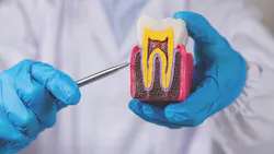
Understanding root resorption
Key Highlights
- Root resorption is a relatively rare but serious dental condition, affecting about 5%–10% of patients and often requiring early identification and specialist referral.
- Two main types—internal and external resorption—differ in origin, clinical presentation, and causes, including trauma, orthodontic forces, infection, or pressure from surrounding structures.
- Advanced imaging such as CBCT improves diagnosis, allowing clinicians to detect smaller lesions and plan appropriate treatment or referral to an endodontist.
The American Association of Endodontists (AAE) has recognized that root resorption is largely considered an endodontic disease affecting the permanent dentition. Naturally, primary dentition is replaced by permanent dentition through root resorption; however, when root resorption occurs in the permanent dentition, it is part of a disease process. It is still not fully understood whether root resorption is due to a physiological process or due to a complex histological action, thus making it difficult to clearly define root resorption.
Generally, root resorption is not a condition that is encountered routinely in dental practices. The occurrence rate is considered to be rare, with approximately 5%–10% of patients experiencing this phenomenon.1 Therefore, reviewing the types of root resorption, clinical presentations, and treatment options is important for early identification, timely referral, and initiation of treatment.
Types of root resorption
The AAE has identified two broad categories of root resorption: internal and external. Distinct differences between both types of resorption have been identified, including resorption site, clinical presentation, and etiology. A more in-depth discussion of each type of root resorption is needed to further understand the differences.
Internal resorption causes advanced destruction of the affected tooth and can involve the adjacent alveolar bone. This type of lesion is inflammatory in nature and is a pulpal disease that involves the resorption of the dentin from the inside of the pulp chamber, affecting the interior walls of the tooth. Internal resorption is often secondary to trauma, can occur as a result of placing a restoration, or can be due to fractures.2 There are three primary types of internal resorption:
Internal inflammatory resorption presents with minor areas of resorption within the root chamber walls. Due to the small size of the lesions, this type of resorption is unable to be detected during an oral examination and often radiographically. If lesions are visible radiographically, they have an oval or balloonlike appearance near the apex.3-5
Internal surface resorption is inflammatory in nature; the lesion occurs in the pulp chamber and can progress through the dentin into the cementum. The etiology of internal surface resorption is commonly associated with trauma or caries.3,4
Internal replacement resorption is very rare. Characteristically, the pulp and dentin undergo replacement by bone. Due to the presence of bone in the root canal, radiographs reveal an oval or balloonlike lesion with a cloudy radiopaque appearance.3-5
Unlike internal resorption, external resorption begins in the precementum lining where the periodontal fibers attach to the root surface. This type of resorption is more common and often is identified in orthodontic patients due to the movement of the teeth.6 The AAE has identified four types of external resorption that have distinct characteristics:
Inflammatory resorption is due to trauma or prolonged trauma to the cementum. The inflammatory component is important because the bacterial infection of the pulp chamber triggers an inflammatory response. Clinically, lesions appear at the apex or along the length of the root. Radiographically this presents as saucer-shaped radiolucent areas along the surface of the root. The etiology of inflammatory resorption can include trauma due to displacement or avulsion.3-5
Replacement resorption is a type of resorption that occurs due to injury to the periodontal ligament or displacement of a tooth. Over time, bone replaces the resorbed area of the root.3-5
Pressure resorption is the result of pressure on the surface of the root. This pressure can be the result of a cyst, tumor, impacted tooth, or rapid orthodontic movement.2-4 It is important to note that when pressure resorption is due to orthodontic treatment, this process is noninflammatory. Radiographically, the roots of teeth appear flat and blunt at the apex.5
Physiological resorption occurs when the primary dentition is being replaced by the permanent dentition.
Identifying root resorption
Root resorption cannot be identified and diagnosed without the assistance of imaging. Subtle clinical changes in tooth color can sometimes be seen in the oral cavity, but this alone cannot be used to make a definitive diagnosis of root resorption. Technical advancements in high-resolution imaging allow for more accurate treatment planning. Resorption can sometimes be identified on traditional periapical radiographs; however, this two-dimensional image has diagnostic limitations.
The use of cone beam computed topography (CBCT) has significantly changed the way dental offices diagnose a variety of conditions, including root resorption. Unlike traditional radiographs, a CBCT is a high-resolution three-dimensional image that allows for better visualization of the oral structures. Resorption lesions can only be seen on traditional radiographs if they are larger than 0.6 mm in diameter.7 In contrast, CBCT imaging can detect smaller root resorption lesions and surrounding tissues by identifying the exact location and size of a lesion. CBCT technology also allows the user to measure the thickness of the tooth and surrounding alveolar bone. This information assists providers in making a thorough and accurate diagnosis of root resorption.
Clinically, patients experiencing resorption may present with vague symptoms that can be attributed to other oral presentations. This is why it is crucial to correlate symptoms with imaging and a complete oral examination. Symptoms of root resorption may include1:
- Pain in any part of the tooth
- Gingival inflammation or swollen tissue
- Increase in the interproximal spaces
- Pink hue in the cervical region of the tooth
- Formation of a “hole” in the tooth, unrelated to caries
Treatment options
Once root resorption has been diagnosed, patients should consult with an endodontist to determine if apical or coronal endodontic treatment is recommended. As with any dental treatment, endodontic treatment is preferred if the tooth can be successfully saved and restored. There are cases of root resorption where the recommendation is to closely monitor the progress or extraction with an implant placement. Generally, cases of internal resorption require careful evaluation to determine restorability, as this condition is associated with a less favorable prognosis. In cases of external resorption, treatment options may vary and can include removal of orthodontic forces, cysts or tumors, endodontic treatment, or extraction if the tooth cannot be preserved. This is why a referral to an endodontist is vital to accurately diagnose and provide treatment recommendations.
Conclusion
Root resorption is a complex condition requiring a multidisciplinary approach to accurately diagnose and treat. Understanding the clinical and radiographic presentations of root resorption will allow dental professionals to identify lesions sooner and make the necessary referrals.
Editor's note: This article appeared in the April/May 2026 print edition of RDH magazine. Dental hygienists in North America are eligible for a complimentary print subscription. Sign up here.
References
- Tooth resorption: internal, external, causes, symptoms, and treatment. Dentaly. Updated April 23, 2025. https://www.dentaly.org/us/oral-health/tooth-resorption/
- Blicher B. Differentiating resorption. American Association of Endodontists. January 5, 2021. https://www.aae.org/specialty/differentiating-resorption/
- Abbott PV, Lin S. Tooth resorption—part 2: a clinical classification. Dent Traumatol. 2022;38(4):267-285. doi:10.1111/edt.12762
- Chandak M Sarangi S, Chaudhari P, Dass A. Revamping the perished: the management of internal tooth resorption. Cureus. 2024;16(5):e61214. doi:10.7759/cureus.61214
- Patel S, Saberi N, Pimental T, Teng PH. Present status and future directions: root resorption. Int Endod J. 2022;55(54):892-921. doi:10.1111/iej.13715
- Koehne T, Zustin J, Amling M, Friderich RE. Radiological and histopathological features of internal tooth resorption. In Vivo. 2020;34(4):1875-1882. doi:10.21873/invivo.11983
- Boas RJEV, Silva RGL, Vaz GR, et al. Root resorption in patients undergoing orthodontic treatment: a literature review. Oral Surg Oral Med Oral Pathol Oral Radiol. 2025;139(5):e142. doi:10.1016/j.oooo.2025.01.682
About the Author

Melissa Van Witzenburg, MS, RDH
Melissa has been practicing dental hygiene for 23 years. She continues to pursue her passion by educating the aging population about oral health and systemic links. Melissa also works clinically in a periodontal office in the Chicagoland area. For more information, email her at [email protected].