Alpha-synuclein in the salivary glands: Clinical significance, pathology, and biomarker potential
Key Highlights
- Alpha-synuclein links oral tissues to Parkinson’s disease: Abnormal α-syn deposits have been identified in salivary glands and other peripheral tissues, supporting the role of the mouth in Parkinson’s research.
- Saliva and oral tissues show diagnostic potential: Salivary gland biopsies, oral mucosal brushings, and saliva testing are being studied as less invasive ways to detect Parkinson’s-related biomarkers.
- Dental professionals can recognize early clues: Xerostomia, drooling, dysphagia, increased plaque accumulation, and oral hygiene challenges may signal underlying neurologic changes and warrant medical referral.
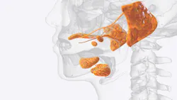
Alpha-synuclein (α-syn) is a small protein best known for its connection to the nervous system and neurodegenerative disease. Under normal conditions, it is found mainly in presynaptic nerve terminals, where it helps regulate synaptic vesicle activity and neurotransmitter release. In simple terms, α-syn helps nerve cells communicate. Although it is most often discussed in relation to the brain, α-syn is also present in peripheral tissues, including the gastrointestinal tract, skin, oral mucosa, and salivary glands. Interest in these peripheral sites has grown because abnormal α-syn aggregation is one of the defining pathologic features of Parkinson’s disease and related synucleinopathies.1
In the salivary glands, α-syn appears to be most clinically relevant within nerve fibers rather than the saliva-producing acinar cells themselves. This distinction matters because salivary secretion depends heavily on autonomic nervous system input, including parasympathetic and sympathetic signaling. Rather than functioning as a simple “salivary protein,” α-syn may serve as a marker of the neural network that helps regulate salivary function. When this network is affected, saliva production, oral clearance, swallowing, and oral comfort may all be disrupted.
In Parkinson’s disease, α-syn can fold incorrectly and build up inside nerve cells. These abnormal protein deposits are a key feature of the disease and may disrupt normal nerve signaling. This process is not limited to the brain. Studies have found abnormal α-syn deposits in peripheral tissues, including the submandibular and minor salivary glands. Beach and colleagues showed that submandibular gland tissue from patients with Parkinson’s disease may contain this pathology, supporting the view that Parkinson’s disease involves both the brain and the peripheral nervous system.2
Can salivary gland biopsies detect Parkinson’s?
The submandibular gland has become one of the most studied salivary gland sites for detecting Parkinson’s-related α-syn pathology. Submandibular gland biopsies have been performed in living patients as part of Parkinson’s disease biomarker studies, including multicenter research evaluating α-syn across saliva, blood, cerebrospinal fluid, skin, colon, and salivary gland tissue. In these studies, gland biopsy has shown diagnostic promise, but it has not yet become a routine clinical test because sensitivity, sampling technique, and tissue adequacy remain important limitations.2,3
Salivary gland biopsy is not a simple screening test. It is more invasive than saliva collection or oral mucosal brushing, requires technical expertise, and can miss pathology if the sampled tissue does not contain enough involved nerve fibers. False negatives are possible and for that reason, salivary gland biopsy should be viewed as an evolving diagnostic tool, not a universal or routine test for every patient with suspected Parkinson’s disease.
Oral brushings: A less invasive diagnostic approach
Because gland biopsy has limitations, researchers are also studying less invasive ways to detect Parkinson’s-related protein changes in the mouth. Cells collected from the oral lining with a small brush have shown abnormal α-syn patterns in people with Parkinson’s disease. This approach is similar in concept to Parkinson’s skin biopsy testing because both look for abnormal α-syn outside the brain, using more accessible body sites as potential biomarkers. The difference is that skin biopsy examines small nerve fibers in the skin and is currently further along clinically, while oral mucosal testing examines cells from the lining of the mouth and remains earlier in development. Still, oral brushing may be easier for patients than gland biopsy or skin punch biopsy, but more research is needed before it can be used in routine clinical practice.4
Saliva as a source of Parkinson’s biomarkers
Saliva is also being studied as a possible diagnostic fluid. Researchers have measured α-syn and other disease-related markers in saliva, including inflammatory proteins and markers linked to neurodegeneration. Recent reviews suggest that certain abnormal forms of α-syn in saliva may be more consistently associated with Parkinson’s disease than total α-syn alone.1,5 This is promising, but the testing methods are not yet standardized enough for chairside diagnosis or routine screening.
What dental professionals should know
For dental professionals, the clinical value is not that we should diagnose Parkinson’s disease from saliva. We should not. The value is that oral findings may contribute to earlier recognition of a broader neurologic picture. Patients with Parkinson’s disease may experience xerostomia, drooling, dysphagia, chewing difficulty, altered taste, increased plaque accumulation, caries risk, periodontal challenges, and difficulty maintaining oral hygiene. Drooling in Parkinson’s disease often reflects impaired swallowing and reduced oral motor clearance rather than true overproduction of saliva. Xerostomia may be related to autonomic dysfunction, medication effects, or reduced salivary flow. These symptoms can significantly affect nutrition, comfort, aspiration risk, and quality of life.
The practical message is clear: the mouth may provide useful clues about systemic neurodegeneration, but the evidence must be interpreted carefully. α-syn pathology in salivary glands supports the concept that Parkinson’s disease is not only a brain disorder; it also affects peripheral neural tissues. Saliva and oral tissues may eventually help with earlier detection or monitoring, but current testing remains investigational. Parkinson’s disease is one of the fastest-growing neurological disorders worldwide, with global prevalence more than doubling over the past 25 years.6 For now, dental providers can recognize patterns in the oral cavity and beyond—unexplained xerostomia, drooling, swallowing difficulty, reduced facial expression, tremor, slowed movement, or loss of smell—and encourage a timely medical evaluation. As research advances, the future is promising, but prudence remains essential.
Editor's note: This article appeared in the August/September 2026 print edition of RDH magazine. Dental hygienists in North America are eligible for a complimentary print subscription. Sign up here.
References
- De Bartolo MI, Belvisi D, Mancinelli R, et al. A systematic review of salivary biomarkers in Parkinson’s disease. Neural Regen Res. 2024;19(12):2613-2625. doi:10.4103/NRR.NRR-D-23-01677
- Beach TG, Adler CH, Dugger BN, et al. Submandibular gland biopsy for the diagnosis of Parkinson disease. J Neuropathol Exp Neurol. 2013;72(2):130-136. doi:10.1097/NEN.0b013e3182805c72
- Chahine LM, Beach TG, Brumm MC, et al. In vivo distribution of α-synuclein in multiple tissues and biofluids in Parkinson disease. Neurology. 2020;95(9):e1267-e1284. doi:10.1212/WNL.0000000000010404
- Zheng Y, Yu Z, Zhao J, et al. Oral mucosa derived α-synuclein as a potential diagnostic biomarker for Parkinson’s disease. Front Aging Neurosci. 2022;14:867528. doi:10.3389/fnagi.2022.867528
- Nijakowski K, Owecki W, Jankowski J, Surdacka A. Salivary biomarkers for Parkinson’s disease: a systematic review with meta-analysis. Cells. 2024;13(4):340. doi:10.3390/cells13040340
- Dorsey ER, Sherer T, Okun MS, Bloem BR. The emerging evidence of the Parkinson pandemic. J Parkinsons Dis. 2018;8(s1):S3-S8. doi:10.3233/JPD-181474
About the Author
 Anne O. Rice, BS, RDH, CDP, FAAOSH
Anne O. Rice, BS, RDH, CDP, FAAOSH
Anne O. Rice, BS, RDH, CDP, FAAOSH, founded Oral Systemic Seminars after over 35 years of clinical practice and is passionate about educating the community on modifiable risk factors for dementia and their relationship to dentistry. She is a certified dementia practitioner, a longevity specialist, a fellow with AAOSH, and has consulted for Weill Cornell Alzheimer’s Prevention Clinic, FAU, and Atria Institute. Reach out to Anne at anneorice.com.