The dental hygienist: Essential to periodontal healing
Key Highlights
- Root cleanliness is central to periodontal healing, with visual confirmation offering advantages over tactile detection alone.
- Periodontal endoscopy and 24% EDTA can improve calculus detection and removal beyond conventional scaling and root planing.
- Addressing the three pillars—root cleanliness, aggressive microorganisms, and occlusion—may help preserve teeth and improve periodontal healing.
The dental hygienist is essential to our periodontal practice. But it wasn’t always that way.
When I left my periodontal residency, I thought I knew what to do in every situation. Pocket elimination was the goal. And perhaps it still is today in many practices. But not for us if our goal is control of chronic periodontitis.
If we look at the forefathers of periodontics, nearly every one of them talks about root cleanliness—how important it is for the root to be clean in order to achieve healing.
We all commonly perform a procedure called scaling and root planing. But that’s a verb. The noun is root cleanliness. What I found over the years is that the more we achieve root cleanliness, the better the opportunity for healing.
Many years ago, I studied with a periodontist, Dr. Perry Ratcliff, in Scottsdale, Arizona. Two of my hygienists and I spent time with him and his hygienists. Dr. Ratcliff’s whole goal was to achieve root cleanliness. He used a retraction cord along with citric acid and tetracycline to give his hygienists better access and improved visibility of the root surface. And sure enough, the patients healed.
I went back to my practice and announced, “We’re going to do the same thing.” At the time, I had a periodontal surgical practice. I decided I was going to stop. I was going to let our hygienists do what Dr. Ratcliff’s hygienists were doing, perform no periodontal surgery other than emergency procedures, and see how the patients healed. If the patients needed surgery, I would do it at no charge. We did that for eight months. In a large periodontal practice, I only had to do four periodontal surgical procedures during that entire period, and they were isolated procedures. I should have stayed with that system.
Periodontal endoscopy
Thirteen years later, we discovered an enhancement to that system: periodontal endoscopy—a camera placed below the gumline, allowing us, for the first time, to visualize calculus. And you know what we found? What we are taught is not necessarily true. A smooth, glassy root, as determined by a no. 17 explorer, doesn’t necessarily accomplish the goal of root cleanliness.
Why? Cobb and Sottosanti describe “fractured calculus.” That means that subgingival calculus will fracture within itself, leaving residual calculus attached to the root surface.¹ And it has been well documented that our effectiveness in achieving root cleanliness with tactile instrumentation is limited in pockets deeper than about 4 mm. Stambaugh and colleagues found that the average depth at which all plaque, calculus, and altered cementum could be removed was only 3.73 mm.²
Once you put a camera into the pocket and see the root surface, what feels smooth often shows calculus.
Every hygienist since that time who has worked in our practice has been humbled to see calculus they had been missing for years. In fact, I know when I have a good hygienist in my practice because he or she comes to me embarrassed, saying, “Look what I have missed all these years.”
Embarrassment isn’t the key. The key is that this is the time for a paradigm shift. If we’re asked to achieve root cleanliness, we need to recognize that visualizing the root is far more beneficial than trying to feel the root.
I’m proud to now have three hygienists who use that periodontal endoscope, place the camera below the gumline, visualize the root surface, clean it, and achieve healing as a result.
But even what we see may not be everything
It was only recently that microislands of calculus were publicized by Drs. Stephen Harrel, Charles Cobb, and their colleagues. Their work demonstrated residual microscopic areas of calculus following scaling and root planing. In a recent study on extracted teeth, conventional SRP was performed with attention to detail using ultrasonic instrumentation and new Gracey curettes, and the procedure was checked with 3.5x loupes. Even after reaching the clinical endpoint of no detectable calculus, 45%–53% of the test area still had residual calculus. Burnishing those surfaces with 24% EDTA gel for one minute reduced the residual calculus to 14%–18%.³
Our procedure for root cleanliness
This is our procedure for root cleanliness:
- Apply 24% EDTA gel to the root surface for one minute using a microbrush and then rinse the EDTA out with an irrigating syringe. This softens the calculus.
- Use the periodontal endoscope to visualize the calculus and remove it using traditional scaling and root planing instruments, including ultrasonics and hand instruments.
- Once we have visual confirmation that the calculus is gone, burnish the root surface again with 24% EDTA gel for one minute using a microbrush.
- Rinse the EDTA out thoroughly.
When this is done thoroughly, the healing can be remarkable.
3 pillars of periodontal healing
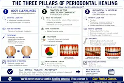
Root cleanliness is one pillar of what I call the Three Pillars of Periodontal Healing.
The second pillar is to determine whether aggressive microorganisms contribute to the disease and, if so, to address them. Watch for what was formerly called “aggressive periodontitis.” What is aggressive periodontitis? It may be referred to as Grade C, but in the current staging and grading system, Grade C refers to rapid destruction.⁴ But when seeing a patient for the first time, how do I know how rapidly the destruction is progressing?
Here’s what I look for:
- The periodontal probe drops into the pocket with no resistance.
- There is no detectable calculus on the root.⁴
I see this in fewer than 10% of my cases, but when it does occur, my protocol changes:
- Identify the bacteria through salivary testing.
- Prescribe the appropriate antibiotic regimen. It is usually amoxicillin and metronidazole.⁵
- Reevaluate the patient for improvement one week after completing the antibiotics.
If I see some clinical improvement, then I feel comfortable moving forward with the periodontal endoscopy protocol.
The third pillar is occlusion. I am looking for fremitus, meaning the tooth moves when the patient is in occlusion. If that happens in a periodontally compromised tooth, the potential for healing is limited. If I see fremitus, I adjust the bite either by removing some tooth structure from the compromised tooth or sometimes adding composite to adjacent teeth so that the occlusal load is shared among several teeth.⁶
Too often, we condemn teeth as “periodontally hopeless” rather than address the three pillars. If we address all three pillars, we can save many more teeth in health and function for many years.
Additional reading: Clinical Tips: Periodontal microscopy and endoscopy
Author’s note: This article was written with the assistance of artificial intelligence.
Editor’s note: This article first appeared in RDH eVillage newsletter, a publication of the Endeavor Business Media Dental Group. Read more articles and subscribe.
References
- Cobb CM, Sottosanti JS. A re-evaluation of scaling and root planing. J Periodontol. 2021;92(10):1370-1378. doi:10.1002/JPER.20-0839
- Stambaugh RV, Dragoo M, Smith DM, Carasali L. The limits of subgingival scaling. Int J Periodontics Restorative Dent. 1981;1(5):30-41.
- Harrel SK, Cobb CM, Nunn ME, Zhao D. Laser and scanning electron microscopy evaluation of residual microislands of calculus. J Periodontol. 2025;96(3):268-278. doi:10.1002/JPER.24-0191
- Harrel SK, Cobb CM, Sottosanti JS, Sheldon LN, Rethman MP. Clinical decisions based on the 2018 Classification of Periodontal Diseases. Compend Contin Educ Dent. 2022;43(1):52-56.
- Sanz M, Herrera D, Kebschull M, et al. Treatment of stage I-III periodontitis—the EFP S3 level clinical practice guideline. J Clin Periodontol. 2020;47(suppl 22):4-60. doi:10.1111/jcpe.13290
- Harrel SK, Nunn ME. The effect of occlusal discrepancies on periodontitis. II. Relationship of occlusal treatment to the progression of periodontal disease. J Periodontol. 2001;72(4):495-505. doi:10.1902/jop.2001.72.4.495
About the Author
 Lee N. Sheldon, DMD
Lee N. Sheldon, DMD
Lee N. Sheldon, DMD, is a periodontist in Melbourne, Florida, and founder of a comprehensive periodontal and restorative dental practice. His clinical interests include periodontal endoscopy, root cleanliness, occlusion, and conservative approaches to saving teeth. He has lectured to dental hygienists and other dental professionals on periodontal healing and treatment planning.