The evolution of the X-ray
Computed radiography will be a familiar tool to the next generation of hygienists, meaning the light box that you`re comfortable with may become a dinosaur.
Robert D. Chadbourne
When Dr. Lawrence Life, a senior partner at Meadow Place Dental, discusses the practice`s decision to incorporate computed radiography, he is very brief about the benefits: "It`s a great team-builder." The decision to toss out the light box in favor of technology that cost $140,000 (every treatment room at Meadow Place Dental was equipped) needed some justification.
"Our approach is to co-diagnose," Dr. Life elaborates. "We help the patient understand his situation and to understand it is his situation; he owns it."
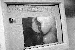
Meadow Dental Place in East Longmeadow, Mass., has10 treatment rooms, manned by three dentists and six hygienists. Computed radiography allows each hygienist and dentist to instantly project a visual or radiographic image onto a screen directly in front of the approximately 6,500 patients who visit the facility. Instead of the cardboard-reinforced film square, a charged coupled device (CCD) or "sensor" is placed in patients` mouths. The sensor transmits through the computer and projects an image within 10 seconds.
"CMOS" technology is the latest enhancement in computed radiography. The image resolution is increased to 12.5 line pairs per millimeter, just about closing the gap with film quality (which has 14). The difference is undetectable to the eye, but CMOS favors imaging when other capabilities are put into play.
Computed radiography systems are in place and part of the curriculum at most of the nation`s 50 dental schools. There has been less effort to get the technology into hygiene schools. Manufacturers generally feel that hygienists can become proficient through on-the-job training. But the subtle marketing to dental students gambles that the future dentist will incorporate computed radiography into the design of an office, perhaps avoiding any package purchase of equipment that includes traditional X-ray systems. Dental schools often obtain price breaks in purchasing such equipment because new dentists tend to buy the equipment they train on; having a product in the teaching facility is a certain way to develop a customer base.
Dr. David Russell, assistant dean of clinical affairs at Tufts University of Dental Medicine in Boston, said, "While new knowledge and new technologies do sometimes originate in teaching facilities, we are not always the first to introduce a piece of equipment. In that vein, I would say it will take a little, but not a lot, more time before the equipment reaches the training programs for hygienists. All schools want to graduate students trained in current technologies, and there is no doubt computed dental radiography is a major advance and is here to stay."
Jan Selwitz, RDH, director of alumni affairs and development at Forsyth Dental Center in Boston, adds, "We`ll be continuing to train in both film and imaging, probably for some years into the future. It will be a long time before all dental offices in the country are switched over."
Forsyth had a single unit installed last summer, leading to a general overview and familiarization program offered during the school`s alumni gathering in October, as well as for some involvement by the entering 55-member class. "They`ll all get an introduction to the unit this year, and there are test questions planned for their block on radiography, but each student may not get a lot of clinical use at the beginning," adds Selwitz.
Computed radiography is an important tool for the dental hygienist because so many of the benefits fall under the umbrella of patient education. Younger patients who have been computer literate for their entire lives can now be more willing participants in preventive dentistry programs. But older patients benefit from the hygienist being able to clearly point out bone density, which reinforces the need for care to ward off periodontic treatment.
Sara Fernald, RDH, a 22-year veteran who works at Meadow Place Dental, was a willing convert. "I had read about the technology, and Dr. Life had really done his homework regarding both the present and upgrading potential for the future," she said.
Fernald believes it is important for hygienists to be proficient in both film and computed radiography to be a fully marketable professional. She said she was able to become proficient with the new technology in less time than she had anticipated, partly because she went through a full-mouth procedure as a patient herself. Fernald estimates that she was comfortable and proficient with the equipment within a week.
After installation of the equipment, she immediately began experiencing better patient education and more extensive patient involvement. "It`s hard to deny what is right in front of you, and it`s easier to get the teaching points across when you`re looking at a 17-inch monitor rather than squinting at a tiny piece of film," Fernald points out.
Projecting dental computer images using digital sensors does not mean the end of the X-ray, but the scorecard of pluses add up in favor of the new systems. Clarity between the two systems is approximately the same, but anything lacking in the computer image is more than offset by the ability to lighten, darken, colorize, measure, or zoom in or out of an area. Size can be amplified 300 times.
X-rays can deteriorate with brown or yellow coloring during storage over a period of time. That is not an issue with computed images, which can be clearly printed onto glossy paper to be sent to a specialist or an insurance company. E-mail transfer of images is not far off into the future.
Storage of images, though, is an issue. The limit is about 7,000 images per gigabyte. "All of this helps if you`re computer-knowledgeable going in," says Dr. Life, adding with a laugh, "One of our hygienists was not, to the extent that she was afraid to touch the mouse for fear she`d break something. Within a week, she was OK."
Perhaps the most rewarding aspect of digital imaging addresses the fact that most hygienists chose their profession because they are "people persons," who rate the interaction with the patient as the most enjoyable aspect of their work. While the setting up and positioning for a digital image takes longer than an X-ray, the overall amount of time spent with computed radiography is less. It also does not require the hygienist to leave the operatory for 10 minutes for full-mouth X-ray development, nor additional time to mount films.
Other issues of importance include:
* Film is flexible, sensors are not, requiring very different patient positioning.
* If an image has been captured incorrectly, it is obvious within a few seconds, and a quick adjustment can be made.
* There are different sizes of sensors for different sizes of patients` mouths, and they are very expensive ($3,000 to $6,000). The "ballpark" figure for outfitting one treatment room is generally about $20,000, a figure that is expected to decrease in the future.
* Extreme care must be taken not to allow metal connections to touch the computer when there is a static charge.
"We all pick up tips, establish our own techniques, and talk back and forth about different situations. This process involves a lot of communication," Dr. Life said. "It looks as if we`ll be saving about $8,000 a year by eliminating mounting, film purchase, the solution, and developing equipment for X-rays. In addition, 90 percent of radiation is eliminated, and environmental considerations, always a matter of concern to hygienists, required to treat X-ray fixers and development fluids and hazardous wastes, are issues of the past."
Experimentation at the most basic level continues. The screens at Meadow Place are wall-mounted, but Dr. Life is not totally satisfied with their height and position. In today`s age of ergonomic planning, optimum treatment room design for both treatment and education need to be part of the evolution into the new approach of computed radiography.
Robert D. Chadbourne is a writer based in East Longmeadow, Mass.
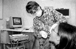
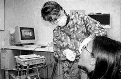
Janet Sessions, RDH, inserts sensor into the mouth of a 12-year-old patient. Photography by Marc Breault.


