Culturally induced pigmented lesions
by Nancy W. Burkhart, RDH, EdD
[email protected]
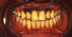
Your patient today is Nadea, a 30-year-old woman from Ethiopia. She has moved to the United States to be with her brother and other family members. This is her first dental visit, and she is being seen in your practice today for radiographs, a new patient dental exam, and a prophylaxis. While reviewing Nadea’s medical history and talking with her, you notice a dark appearance of the maxillary gingiva when she smiles (see Figure 1).
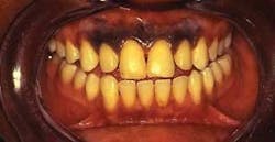
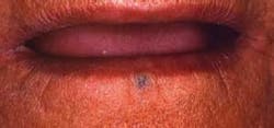
Figure 1 (above) and Figure 2 (below): Both images were contributed by Dr. Doron Aframian and appear in General and Oral Pathology for the Dental Hygienist (Lippincott, Williams and Wilkins).
Mentally, you make a note to ask about this dark area when you begin your intraoral exam. Since she has a very dark complexion, your first instinct is to attribute the dark pigmentation to racial pigmentation. As you question Nadea, she tells you that the pigmentation has been present for as long as she can remember, and most of the female family members in her Ethiopian community have the same type of pigmentation. Nadea reports that the gingiva of young children is routinely darkened at a very young age, using a technique of abrasion and then rubbing soot/herbs into the tissue.
Certain cultural practices use pigmentation of the tissues to depict beauty, promote health, denote right of passage, and to fulfill various functions such as warding off bad spirits and promoting good luck.
Etiology: Chronic and long-term injury with assault to the tissue results in inflammatory pigmentation. Some medications such as minocycline, antimalarials, oral contraceptives, and thermal changes may cause pigmentation as well. Pigmentation may be present due to diseases such as Peutz-Jeghers syndrome, Addison’s disease, Kaposi’s sarcoma, Laugier-Hunziker syndrome, and melanoma.
Physiological pigmentation is caused by the tendency to produce melanin in varying amounts. An overproduction of melanin occurs in racial pigmentation, and these areas will not blanch when pressure is applied. Pigmentation sometimes occurs in hormone therapy and hormone-production states such as those seen in pregnancy. Additionally, long-term inflammation in the tissues due to disorders such as lichen planus will cause increased pigmentation, and long-term use of tobacco products can produce smoker’s melanosis.
In the case presented here, the pigmentation is induced by trauma and dark pigments. Nadea tells you that it is a custom performed in her community. The gingiva is abraded and black soot (sometimes a coloring agent called lampblack) is placed in the tissue to promote a dark pigmentation. Typically, the tattooing is confined to the canines for males and is more extensive for females covering the gingival surface from canine to canine as depicted in Figure 1. Multiple attempts are made to cause abrasion of the tissues with the layering of the soot material until the desired pigmentation is accomplished.
Additional tissue pigmentation is reported throughout the literature with regard to other types of beauty and oral enhancements in various cultures. Figure 2 depicts a woman from Arabia with an ethnic tattoo below the lower lip. This tattoo was performed in her culture to ward off bad luck.
In cases presented by Rawal et al., a patient reported having the placement of pigments performed at age six with a second placement at age 10. The authors also report a woman from Dakar, Senegal, having pigments placed at age 40 in a dental office. The patient decided to have the procedure much later in life since she did not have the customary pigmentation early in childhood. Those performing cultural tattooing from the woman’s community brought in the pigments that could be placed while she had anesthesia administered. A strong cultural propensity to adapt to community standards remains, promoting the ethnic beliefs of those around them.
Discussion: Racial pigmentation and disease states may be confused with gingival tattoos, but the astute clinician can compare the color variations of the tissues and question the patient about previous cultural tattooing. Unintentional tattooing can result from amalgam fragments (focal argyrosis), pencil injuries with graphite fragments, and dust from metal fragments due to dental burs or metal particles. Radiographs may confirm metal fragments rendering a diagnosis of metals within the tissues when this is expected. In the case of imbedded herbs, soot, and plant-based pigments, no radiopaque implications would be observed.
Conclusions: Careful assessment of extraoral tissues and any pigmented tissue intraorally should be evaluated. Any pigmented lesions should have a determined etiology. Possible biopsy may occur if the tattoo is mistaken for serious disease states such as melanoma.
With that said, the reverse may be true as well with misdiagnosis and a failure to recognize a more serious type of disease. The United States has become truly multicultural with many communities/ethnic groups appearing in all areas of the country. The dental clinician should be aware of cultural practices that may be evident during the dental exam and be able to ask relevant questions in making assessments. Listen to your patients and keep asking good questions.
References
DeLong L, Burkhart N. General and Oral Pathology for the Dental Hygienist, Lippincott, Williams and Wilkins, Baltimore, Maryland, 2008.
Mani NJ. Gingival Tattoo. A hitherto undescribed mucosal pigmentation. Quintessence Int.1985;16:157-159.
Rawal SY, Burrell R, Hamidi CS, Kalmar JR, Tatakis DN. Diffuse pigmentation of maxillary attached gingiva: Four cases of the cultural practice of gingival tattoo. J Periodontol.78:1, 2007.
Regezi JA, Sciubba JJ, Jordan RCK. Pigmented lesions. In: Regezi JA, Sciubba JJ. Jordan RCK, eds. Oral Pathology: Clinical Pathologic Correlations, 4th ed. St. Louis: Saunders: 2003:129-142.
Nancy W. Burkhart, BSDH, EdD, is an adjunct associate professor in the department of periodontics, Baylor College of Dentistry and the Texas A & M Health Science Center, Dallas. Dr. Burkhart is founder and co-host of the International Oral Lichen Planus Support Group http://www.bcd.tamhsc.edu/outreach/lichen/ and coauthor of General and Oral Pathology for the Dental Hygienist. Her Web site for seminars is www.nancywburkhart.com.