Case Study: Case #1
A 16-year-old female visited a dental office for a routine check-up and prophylaxis. Radiographic examination revealed a tooth-like radiopacity between the maxillary central incisors.
null
History
The patient denied any history of signs or symptoms associated with the maxillary central incisor region. The patient appeared to be in a general good state of health, with no significant medical history.
The patient’s dental history included regular dental examinations and routine dental treatment. At the time of the dental appointment, the patient was not taking medications of any kind.
Examinations
The patient’s vital signs were all found to be within normal limits. Examination of the head and neck region revealed no enlarged or palpable lymph nodes. Examination of the oral soft tissues revealed no unusual findings. No bony abnormalities were noted. Examination of the teeth revealed a large diastema between the maxillary central incisors.
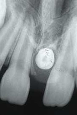
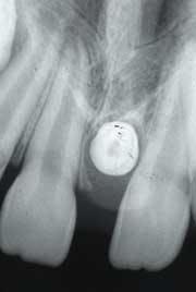
After a thorough clinical examination, four bite-wing radiographs and a panoramic film were ordered. In addition, a periapical film was ordered to examine the diastema area between the maxillary central incisors. Examination of the periapical radiograph revealed a dense tooth-like radiopacity surrounded by a radiolucent rim (see radiograph). The radiopacity was also apparent on the panoramic film.
Following the radiographic examination, the teeth adjacent to the lesion were pulp tested for vitality with an electric pulp tester; both incisors tested vital. No expansion or bony changes were noted in the area of the lesion.
Clinical diagnosis
Based on the clinical and radiographic information available, which of the following is the most likely diagnosis?
º compound odontoma
º ossifying fibroma
º mesiodens
º osteoma
º benign cementoblastoma
Diagnosis
º mesiodens
Discussion
The mesiodens is a supernumerary or “extra” tooth. (The term supernumerary means in excess of the normal or regular number.) The mesiodens is developmental in origin and is comprised of enamel, dentin, cementum, and pulp. The mesiodens may occur in the deciduous or permanent dentitions, although the permanent dentition is affected more frequently.
Only a small percentage of the population exhibits supernumerary teeth. The most common supernumerary tooth is the mesiodens; the second most common supernumerary tooth is the distomolar. Supernumerary teeth can be found in any location.
Clinical features
The mesiodens is a smaller than normal sized tooth that is found at the midline of the maxilla. (Mesio refers to midline, dens refers to tooth.) The shape of the mesiodens may resemble a normal tooth or exhibit a conical-shaped crown and a short root. The mesiodens may erupt and be evident clinically, or remain embedded or impacted; most mesiodens do not erupt. When impacted, the mesiodens is often situated in an inverted position. The mesiodens may occur singly or in multiples and is asymptomatic.
Radiographic features
The mesiodens is typically discovered during radiographic examination. The dental radiograph is useful in identifying the appearance, location, and number of unerupted supernumerary teeth. The mesiodens is easily viewed on periapical, occlusal, or panoramic radiographs. Supernumerary teeth, like the mesiodens, can be detected on dental radiographs in the primary dentition after the age of three or four, and, in the permanent dentition after the ages of nine to 12.
As viewed on a dental radiograph, the mesiodens appears as a tooth-like radiopacity. This tooth-like radiopacity may appear rudimentary or conical in shape, or, resemble teeth normally found in the region. The size may vary from normal to miniature.
Diagnosis
The diagnosis of a mesiodens is based on its radiographic appearance. On a good quality radiograph, the outline of a tooth and the radiolucent shadows of the pulp cavity and surrounding follicle are characteristic for the mesiodens.
Treatment
The treatment for the mesiodens depends on the location, position, and number of teeth, and the complications that may arise from surgical removal. The unerupted mesiodens is often associated with cyst development, root resorption and the impediment of tooth eruption. In such cases, surgical removal is indicated.
The erupted mesiodens is non-functional and may cause complications with the eruption of normal teeth, or, cause crowding and mal-positioning of the adjacent teeth. The erupted mesiodens should be extracted. In instances where a patient exhibits multiple supernumerary teeth, conditions such as Gardner’s syndrome and cleidocranial dysplasia must be ruled out.
Joen Iannucci Haring, DDS, MS, is a professor of clinical dentistry, Section of Primary Care, The Ohio State University College of Dentistry.