What are the implications of residual root sockets?
Dental professionals may evaluate or treat patients who complain of pain in areas where a tooth has been extracted, and often there are no clinical signs of any disease process in the mucosal tissue. The pain in question could even be in a totally edentulous area where the extractions occurred many years prior to the complaint of pain. This can be very confusing for both the patient and the clinician. When we think of the causes of oral pain, the reason is tooth-related most of the time.
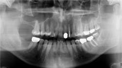
When a radiograph is taken, the dental professional may see what appears to be the outline of the root socket where an extraction has occurred, usually many years prior to any complaints (figures 1 and 2). This is a residual socket or residual root socket. The outline of the tooth’s root cavity is faintly visible, and when examined surgically, it may be hollow or filled with dense fibrous scar tissue, granulation tissue, or very immature bone.
Residual root sockets have also been referred to as ghost sockets or laminar rain. These have been associated with poor blood flow or postsurgical sepsis and continuing or chronic infection of the wound site. With residual root sockets, pain is sometimes present with no clinical signs of infection, making it somewhat perplexing to the dental professional. The concept was introduced in the 1930s but is not part of the then-popular theory of classic residual infection, which proposed these could cause systemic diseases. “Residual root socket” merely refers to an area of alveolar bone that was not able to heal. As stated in 2012 by Bouquot et al., this “is evidence of a mild, chronically diseased alveolar bone (mostly ischemic, sometimes inflammatory) and the underlying bone condition remains indefinitely unless the bone is treated.”1 It does not imply disease spread to the bloodstream or beyond the jaws.
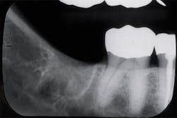
Figure 1: Residual sockets. Note the outline of the previous roots of the molar tooth. Courtesy of Jerry E. Bouquot, DDS, MSD, DABOMP, DABOM (hon), FAAOMP, FICD, FACD, FRCM (UK), emeritus professor and past chair, Department of Diagnostic and Biomedical Sciences, University of Texas, Houston, Texas.
A residual root socket may be seen on a radiograph and is typically a nonurgent phenomenon unless the patient calls attention to the specific area due to discomfort or pain. Since the initial reports on residual root sockets, we have a much better understanding of bone health and healing, along with the intricate nature of bone development and loss. Implant surgery, in particular, has become widely accepted and successful. It is not known whether placing implants in bone with radiographic evidence of a residual root socket is contraindicated.
In a 2012 article about ghost roots, Bouquot et al. point out that these also show faint root shapes but are actually poorly formed dental roots, not bone lesions.2 More commonly, the entire tooth is involved and is then called a ghost tooth. Such teeth are characteristically associated with regional odontodysplasia. The teeth are so poorly formed that they only appear as faint shadows of teeth. A residual root socket is an independent finding, and the tooth is no longer in the socket.2
In most patients with extractions, bone fills in the defect over time. Why some individuals develop residual root sockets and never remodel bone in extraction sites is unknown, although it is thought that perhaps bacterial or viral contamination during removal might be involved in this process.
Poor blood flow might also be a factor. It is known that older individuals regularly show evidence of inadequate blood flow with reductions in vascular supply and a diminished amount of radiographically “normal” edentulous alveolar bone. Many oral surgeons removing teeth for implants and for dentures will have patients take pentoxifylline (Trental 400) for two weeks prior to and after surgery to improve circulation to the areas where extractions will be made. The goal is to increase the blood supply to the tooth areas, and recent reports suggest that Trental assists in promoting wound healing.3 A healthy, adequate blood supply is known to normalize pH in the body and provide cellularity, rapid growth, and remodeling and repair of bone and tissues.
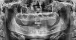
Figure 2: Residual sockets in the same left mandible. Courtesy of Dr. Jerry E. Bouquot.
Bone health is important and crucial in removing waste products and combating acid in the body. The vascular supply decreases with age and is especially an issue in the elderly. In older individuals, there may be regions of asymptomatic chronic ischemic bone disease and osteonecrosis. It is well known that certain disease states affect bone loss and vascular supply to bone and tissues, including diabetes, anemia, chronic airway diseases, and immobility. Some lifestyle habits—such as smoking, due to the nicotine, which is a vasoconstrictor—also contribute to bone loss, according to Marenzana and Arnett.4 Osteoporosis is another limiting factor in bone health within the older population. Marenzana and Arnett also mention that doppler ultrasound measurements showed that femoral artery blood flow was about 30% lower in older men (average age 64 years), compared with younger men (average age 28 years), whereas vascular resistance was about 50% higher.4
Figure 3 shows a patient in his 60s who had a third-molar extraction. He developed an 11 mm pocket on the distal of No. 31 after the extraction of No. 32 and never regained the bone associated with this area. The patient had no pain, but the tooth did have mobility. The surgeon waited to assess the bone in the distal area of No. 31, but since the area never filled in, the tooth was eventually extracted. This depicts an example of bone loss that was never regained or filled in with healthy bone.
Even though most areas with residual root sockets remain trouble-free, there are patients who present with pain years after extractions have occurred. In a 2012 article, Bouquet et al. recommend that “when pain is involved, additional testing with scintigraph, quantitative ultrasound, diagnostic anesthesia, and surgical exploration is recommended.”1 They write, “This is especially important because previous follow-up studies of such areas, with or without [residual root sockets], have shown that surgical curettage of ischemic or inflammatory bone or marrow eliminates the pain, indefinitely, for 72%–86% of treated patients.”1

Figure 3: Chronic bone loss in the area of No. 31. Extraction occurred without subsequent bone formation. Courtesy of Carol Perkins, BA, RDH.
Hygienists are constantly involved in taking and evaluating radiographs. Sometimes dental complaints and soft-tissue problems are found during recare appointments and might or might not be called to the dental professional’s attention by the patient. Intact surface tissue, without ulcerations or any indications of a problem, may be misleading . . . the problem could be deep in the bone.
This column is intended to call attention to extraction sites with residual root sockets that may be viewed on radiographs with or without a complaint by the patient. Careful documentation, possible referral, and follow-up are indicated. If the patient is complaining of pain or tenderness, even though a tooth is not present, careful evaluation of the radiograph and bone area is warranted.
As always, continue to listen to your patients and always ask good questions.
References
1. Bouquot JE, Glaros WP, Zarghouni S. Residual sockets: Radiographic, surgical, microscopic features in 76 cases. Oral Surg Oral Med Oral Pathol Oral Radiol. 2012;114(3):e61-e62.
2. Bouquot JE, Dorn MS, Huynh C. Ghost roots: The strangest roots of all? J Gt Houst Dent Soc. 2012;84:15-17.
3. Ahmadi M, Khalili H. Potential benefits of pentoxifylline on wound healing. Expert Rev Clin Pharmacol. 2016;9(1):129-142. doi:10.1586/17512433.2016.1109443.
4. Marenzana M, Arnett TR. The key role of the blood supply to bone. Bone Res. 2013;1(3):203-215. doi:10.4248/BR201303001.
About the Author

Nancy W. Burkhart, EdD, MEd, BSDH, AAFAAOM
NANCY W. BURKHART, EdD, MEd, BSDH, AAFAAOM, is an adjunct professor in the Department of Periodontics-Stomatology, College of Dentistry, Texas A&M University, Dallas, Texas. She is founder and cohost of the International Oral Lichen Planus Support Group (dentistry.tamhsc.edu/olp/) and coauthor of General and Oral Pathology for the Dental Hygienist, now in its third edition. Dr. Burkhart is an academic affiliate fellow with the American Academy of Oral Medicine, where she also serves as chair of the Affiliate Fellowship Program Committee. She was awarded the Dental Professional of the Year in 2017 through the International Pemphigus and Pemphigoid Foundation, and she is a 2017 Sunstar/RDH Award of Distinction recipient. Her professional interests are in the areas of oral medicine and the relationship between oral and whole-body health, with a focus on mucosal disease and early oral cancer detection. Contact her at [email protected].