A 27-year-old male visited a dentists office for treatment of multiple painful oral lesions
A 27-year-old male visited a dentist`s office for treatment of multiple painful oral lesions and crusted lips.
Joen Iannucci Haring, DDS, MS
History
The patient stated that the sores in his mouth had started abruptly four days earlier. He described the sores as painful and claimed that both eating and drinking intensified the pain. Since the onset, the lesions had increased in number and the pain had worsened. When questioned about constitutional symptoms, the patient claimed an overall feeling of malaise, headache, and a low-grade fever. The patient denied any history of a previous episode.
The patient`s medical history was reviewed. No significant findings were noted, with the exception of a Herpes simplex viral infection approximately two weeks earlier. At the time of the examination, the patient was taking Tylenol for the oral pain.
Examinations
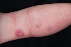
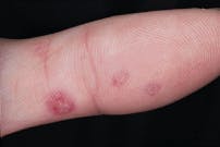
Examination of the head and neck regions revealed enlarged superficial cervical and submandibular lymph nodes. No skin lesions were apparent in the head and neck regions. Examination of the skin on the hands and arms revealed multiple scattered vesicles surrounded by ring-like areas of redness (see photo). Oral examination revealed multiple ulcerations scattered throughout the oral cavity and a hemorrhagic crusting of the lips (see photo).
Differential diagnosis
Based on the clinical information provided, which one of the following is the most likely diagnosis?
- Benign mucous membrane pemphigoid
- Pemphigus vulgaris
- Erythema multiforme
- Primary herpetic gingivostomatitis
- Herpetiform aphthous ulcers
Diagnosis
- Erythema multiforme
Discussion
Erythema multiforme is an acute vesiculobullous disease that affects the skin, the oral mucous membranes, or both. The exact cause of erythema multiforme (EM) is unknown, although an immunologically mediated process is suspected. A recent infection or drug is often implicated as a triggering factor for this disease. In approximately 50 percent of cases, one of the following is implicated:
- A preceding infection with the Herpes simplex virus
- Mycoplasma pneumonia
- The recent use of a medication, particularly an antibiotic or analgesic.
The most common precipitating factor is a Herpes simplex viral infection, preceding the onset of EM by one to three weeks.
Clinical features
Erythema multiforme is a disease that occurs in young adults ages 20 to 30, although it can affect any age group. Males are affected more frequently than females. This disease has a sudden, sometimes explosive onset and runs a variable course of two to six weeks. Recurrences are not uncommon; EM may recur seasonally in the spring and fall. Prodromal signs and symptoms occur approximately one week prior to onset and include fever, malaise, and headache.
The term erythema multiforme is descriptive of this disease, specifically the skin lesions. The skin lesions that occur with EM appear in many forms (for example, macules, papules, vesicles, and bullae), hence the term multiforme. The term erythema refers to the red appearance of these lesions.
The skin lesions seen with EM have a classic appearance and are known as target lesions. The concentric erythematous pattern of the skin lesions is said to resemble a target or a bull?s eye. Clinically, the target lesions appear as round macules; each macule is made up of concentric red rings that are separated by rings of normal skin color. The center of each round macule may be pale, vesicular, bullous, or ulcerated. Target lesions range in size from several millimeters to several centimeters in diameter. These skin lesions develop in approximately one-half of the cases of EM and are usually seen on the skin of the arms, hands, wrists, legs, and ankles. In some cases, the distribution appears symmetric.
The oral lesions that occur with EM are vesiculobullous in nature; when the short-lived vesicles and bullae rupture, ulceration results. The ulcerations seen with EM may vary in number and clinical presentation. The number of lesions seen may vary from a few scattered ulcerations to multiple lesions involving the entire oral cavity. Any area of the oral cavity may be involved. The lips are most consistently affected with multiple, painful, hemorrhagic, and crusting ulcerations and erosions. The labial and buccal mucosa, hard and soft palates, and tongue may be involved. The intraoral ulcerations and raw erosions exhibit mucosal sloughing and bleed freely. A diffuse redness of the oral mucosa may be present. Depending on the severity of the oral involvement, the symptoms may range from a mild discomfort to a marked, severe pain. Systemic manifestations such as fever, malaise, headache, and lymphadenopathy are often present.
Diagnosis
EM is diagnosed on a clinical basis. The diagnosis is established based on the clinical signs, symptoms, and patient history. In the absence of the characteristic skin lesions, it may be difficult to make the diagnosis of EM. A thorough patient history in regards to similar attacks and previous skin lesions may be helpful in making the clinical diagnosis. EM does not have a unique histologic appearance and therefore cannot be diagnosed via biopsy and microscopic examination. Immunopathologic studies are not specific for EM as well. The diagnosis is essentially one of exclusion and is made after all other possibilities (vesiculobullous diseases, for example) have been ruled out.
Treatment
Management of a severe case of EM includes the use of systemic corticosteroid medications, especially in the early approximately one week prior to onset and include fever, malaise, and headache.
The term erythema multiforme is descriptive of this disease, specifically the skin lesions. The skin lesions that occur with EM appear in many forms (for example, macules, papules, vesicles, and bullae), hence the term multiforme. The term erythema refers to the red appearance of these lesions.
The skin lesions seen with EM have a classic appearance and are known as target lesions. The concentric erythematous pattern of the skin lesions is said to resemble a target or a bull?s eye. Clinically, the target lesions appear as round macules; each macule is made up of concentric red rings that are separated by rings of normal skin color. The center of each round macule may be pale, vesicular, bullous, or ulcerated. Target lesions range in size from several millimeters to several centimeters in diameter. These skin lesions develop in approximately one-half of the cases of EM and are usually seen on the skin of the arms, hands, wrists, legs, and ankles. In some cases, the distribution appears symmetric.
The oral lesions that occur with EM are vesiculobullous in nature; when the short-lived vesicles and bullae rupture, ulceration results. The ulcerations seen with EM may vary in number and clinical presentation. The number of lesions seen may vary from a few scattered ulcerations to multiple lesions involving the entire oral cavity. Any area of the oral cavity may be involved. The lips are most consistently affected with multiple, painful, hemorrhagic, and crusting ulcerations and erosions. The labial and buccal mucosa, hard and soft palates, and tongue may be involved. The intraoral ulcerations and raw erosions exhibit mucosal sloughing and bleed freely. A diffuse redness of the oral mucosa may be present. Depending on the severity of the oral involvement, the symptoms may range from a mild discomfort to a marked, severe pain. Systemic manifestations such as fever, malaise, headache, and lymphadenopathy are often present.
Diagnosis
EM is diagnosed on a clinical basis. The diagnosis is established based on the clinical signs, symptoms, and patient history. In the absence of the characteristic skin lesions, it may be difficult to make the diagnosis of EM. A thorough patient history in regards to similar attacks and previous skin lesions may be helpful in making the clinical diagnosis. EM does not have a unique histologic appearance and therefore cannot be diagnosed via biopsy and microscopic examination. Immunopathologic studies are not specific for EM as well. The diagnosis is essentially one of exclusion and is made after all other possibilities (vesiculobullous diseases, for example) have been ruled out.
Treatment
Management of a severe case of EM includes the use of systemic corticosteroid medications, especially in the early stages of the disease. Less severe cases of EM can be treated with topical corticosteroid rinses. Other treatments include symptomatic and supportive therapies. Analgesics/antipyretics can be used to reduce pain and fever and alleviate the aches and pains of malaise. An adequate fluid intake is important to prevent dehydration. Palliative oral rinses can be used to lessen the discomfort of the oral lesions.
In cases where recurrent episodes of EM are a problem, initiating factors such as a recent Herpes simplex infection or drug exposure should be considered. In such cases of Herpes simplex infections, continuous acyclovir therapy may be used to prevent such recurrences. In cases of a known drug exposure, the precipitating drug must be avoided.
Joen Iannucci Haring, DDS, MS, is an associate professor of clinical dentistry, Section of Primary Care, The Ohio State University College of Dentistry.

