The Golden Rule
Julie believes in the Golden Rule now more than ever. “Do unto others as you would have them do unto you” is endorsed by all the great world religions. Many of us take this message to heart in our personal relationships, try to live this way, and teach our children this way of life.
As a dental hygienist, are you practicing the Golden Rule by performing a complete soft-tissue oral cancer evaluation for every patient? It only takes five minutes to perform a thorough manual and visual head and neck soft-tissue oral cancer screening. How about you, when were you last checked?
Julie’s story
Julie is a practicing dental hygienist and owner of a promotions company. Three years ago she was doing a head and neck exam on herself and found a pea-sized lump behind her right ear. At first she was dumbfounded and thought, “What is that?” For days she checked and rechecked and went crazy analyzing what it could be. She finally went to her physician to show him the lump. The physician gave her an antibiotic to see if it would go away. It didn’t.
Julie kept checking the lump constantly. As the mother of three small children, she was overcome with worry. Julie took the bull by the horns and visited a well-known oral surgeon. She asked for a panorex so she could look at the lump herself. The surgeon tried to reassure her by telling her it was probably nothing, and she didn’t need to worry because it didn’t show up on the film. If she had been like many patients, this response would have been great news and she would have gone on her way.
Yet Julie is a hygienist and tends to analyze; some people even call it overanalyzing. She could not accept the oral surgeon’s response. She just couldn’t walk away because she felt there was more. Something deep inside her told her to explore further. She decided to visit the otolaryngologist (ENT) who had removed her tonsils when she was 20. The ENT tested using a fine needle biopsy, which revealed a tumor of Julie’s parotid gland. The ENT thought a simple in-office superficial parotidectomy would be sufficient. After a few days he called to say removal would require more than an in-office procedure. Something in what he said told Julie he wasn’t very familiar with the procedure she needed, and she knew she needed more information. She decided to get out her dental hygiene books and search the Web. She had to keep ownership of her health, and before surgery she wanted to know more.
On her first search of the Web, she found many people who had had the procedure done. She viewed pictures and read descriptions that made her more fearful.
Julie searches for answers
As a hygienist, Julie knew the parotid gland is a major salivary gland. She knew these glands are located in front of the ears, but she had forgotten how far they extend to the area beneath the earlobe along the lower border of the mandible.
She learned a few basic principles about salivary gland tumors: the larger the salivary gland, the less likely the tumor will be malignant. Thus, a mass in the parotid has only a 25 percent chance of being a malignant tumor, whereas a mass in the sublingual gland has a 75 percent chance of being a malignant tumor. Masses in the submandibular gland have about a 50 percent chance of being malignant. The method of choice for evaluating a parotid mass is a fine needle biopsy.Julie was glad she sought out her ENT for the evaluation because the diagnostics went beyond a panorex.
Although most tumors grow slowly and are benign, they can continue to grow and become malignant. Surgical removal later may be more difficult. In general, any lump in front of or below the ear must be considered a parotid mass until proven otherwise. A fine needle biopsy can confirm that a tumor is cancerous but not if it is benign. Usually, parotid masses are resected with a superficial parotidectomy. This is for two reasons: ➊ It is very easy to damage the facial nerve unless it is traced out from its origin throughout the entire course in the gland. ➋ The most common types of salivary tumors tend to recur. This allows medical personnel to get a good margin of tissue around it, and hopefully decrease the recurrence rate.
Julie was concerned because she didn’t think her ENT knew much about the surgery she needed. She didn’t want to risk permanent damage, and she wondered if there could be other side effects from surgery. Her ENT admitted this was not his specialty and asked Julie to visit someone else in the group. She appreciated his honesty.

The next ENT reviewed Julie’s pathology reports, panorex, and data. Julie requested another biopsy. Rather than performing this himself, he sent Julie to a well-respected pathologist who was very reassuring and told her he was going to get samples from three sites to make a complete diagnosis. The results of the second biopsy were the same. It was a benign parotid tumor that was slowly growing. Plans were set to remove it.
Julie’s parotidectomy took more than four hours. Two surgeons performed the procedure together. One surgeon was there for the sole purpose of monitoring Julie’s facial nerve. She stayed in the hospital three days while her neck and ear area drained. She received 22 stitches from the top of her front ear down her neck. The ENT said he was very glad the tumor was out. It had grown to the size of a golf ball and had started growing around her neck muscles, much to everyone’s surprise.
Julie’s saga doesn’t end here
Julie went for her annual exam expecting no problems. She had received a clean bill of health year after year. But this time she was told she needed a biopsy because her Pap test had come back with positive results. In just one year she had gone from normal to positive. At only 36 years old, Julie was in shock. She knew nothing about cervical cancer, such as how common or treatable it is if found early. She was simply very grateful for these types of tests for this common cancer and precancerous cells.
Julie’s outpatient surgery was successful. She was given a clean bill of health, but must undergo more frequent Pap tests. While she feels great, her experiences have led her to think differently about what hygienists do in dental practices.
What if Julie hadn’t had a Pap smear? She had no risk factors. Just like we do in dentistry, the gynecologist could have handed her a list of precautions to watch. The list would include:
✰ A sore that bleeds easily or does not heal
✰ A color change of the tissues
✰A lump, thickening, rough spot, crust or small eroded area
✰ Pain, tenderness, or numbness anywhere
This list of late stage manifestations is from a popular oral cancer brochure. Should Julie have waited and watched? Should our patients wait and watch?
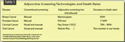
Many women have an annual Pap test along with a manual and visual exam. Women undergo the exam because they know the importance of early cervical cancer detection. According to the American Cancer Society, approximately 55 million Pap smears are performed each year in the United States. Early detection of cervical cancer has increased the five-year survival rate by approximately 91 percent. Women ages 35 to 55 are considered at highest risk for cervical cancer. Table 1 shows how adjunctive screening technologies have made a difference.
The statistics for oral cancer have not decreased in over 30 years. Though a majority of dental professionals claim to screen for oral cancer, an American Dental Association survey said only 15 percent of patients realized they had undergone an exam. What does screening mean? Stedman’s medical dictionary defines screening as, “The examination of a group of usually asymptomatic individuals to detect those with a high probability of having or developing a given disease.”
The first step is the manual and visual oral cancer screening every hygienist learned in school. Yet, we can do more. ViziLite Plus with TBlue oral lesion identifying and marking system (Zila Pharmaceuticals, www.vizilite.com, Phoenix, Ariz.) is the only FDA-approved technology for adjunctive screening of oral abnormalities that could lead to oral cancer. Conventional visualization with incandescent illumination provides identification of only well-developed surface abnormalities. With ViziLite Plus, lesions can be identified prior to visualization.
ViziLite Plus with TBlue works because the density of the nuclear content and mitochondrial matrix of abnormal cells is typically greater than normal cells. The increased molecular density may reveal the increased proliferative rate and metabolic activity of precancerous cells. The ViziLite exam helps examiners see the difference in the nuclear/cytoplasmic ratio of dysplastic cells. After rinsing with a dilute acetic acid solution, the dense nucleus of abnormal squamous epithelium tissue appears white when viewed under a diffuse, low-energy wavelength light. Normal epithelium absorbs the light and appears dark. ViziLite Plus can identify an abnormality. Lesions stained with TBlue can be viewed clearly for an intraoral photo to include in the patient record, submit to an insurance carrier, or refer to a specialist.
Our patients rely on us to be the experts in oral health. We’re their best defense against oral cancer. We are, in fact, lifesavers. To Julie, it isn’t just about adding another tool to her armamentarium. It’s about practicing the Golden Rule. She knows the difference early screening can make. She knows cells can change at any time. She knows there are slow-growing lesions or rapidly changing cells, and she realizes no one can see and feel everything. Julie wants all professional practitioners to look at how they can make a difference. She challenges each of us to enhance our standard of care.
References
1 Parotid gland surgery, Mayo Clinic, www.mayoclinic.org/salivaryglandtumors/parotid.html. Accessed 1/2/06.
2 Staffell J. Salivary gland disease. University of Texas Health Science Center, www.uthscsa.edu/oto/salivary.html. Accessed 1/2/06.
3 Smith R, Mettlin C, Davis K, Erye H. American cancer society guidelines for the early detection of cancer. American Cancer Society, http://www.cancer.org/docroot/PUB/content/PUB_3_8X_American_Cancer_Society_Guidelines_for_the_Early_Detection_of_Cancer_update_2001.asp. Accessed 1/8/06.
4 Questions and answers about cigar smoking and cancer. (2000) U.S. Department of Health and Human Services, National Institutes of Health, National Cancer Institute, http://cis.nci.nih.gov/fact/10_16.htm. Accessed 9/1/05.
5 The importance of early detection. American Dental Association, http://www.ada.org/public/topics/cancer_oral.asp. Accessed 12/31/05.
6 Medical malpractice litigation: head and neck cancer. (2004) American Academy of Otolaryngology, www.thedoctors.com/pdf/riskmanagement/aaohnspracmgmt.pdf. Accessed 1/2/06.
7 Lydiatt DD. Cancer of the oral cavity and medical malpractice. Laryngoscope 2002; (112):816-819.