Case #3
by Joen Iannucci Haring
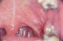
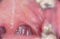
A 25-year-old female visited her dentist for a routine checkup. During the examination, a mass on the soft palate was noted.
History
The patient was unaware of the mass on the soft palate. The patient denied any pain or discomfort associated with the lesion. The patient did not recall any history of trauma to the affected area.
During the dental appointment, the patient appeared to be in an overall good state of health with no significant medical history. The patient was not taking medications of any kind. Past dental history included dental examinations at irregular intervals and routine dental treatment. The patient's last dental appointment was six months earlier for a dental prophylaxis.
Examinations
Physical examination of the head and neck regions revealed no enlarged or palpable lymph nodes. The patient's vital signs were all within normal limits. No significant extraoral findings were noted.
Intraoral examination revealed a pale pink mass on the soft palate. The lesion appeared to measure less than one centimeter in diameter and felt firm when palpated. A smooth surface texture without ulceration was noted. Further examination of the oral soft tissues revealed no other masses present.
The patient was referred to an oral surgeon for an excisional biopsy and surgical removal of the lesion. Histologic examination of the tissue revealed epithelial cells in a nest-like arrangement with pools of myxoid, chondroid, and mucoid material.
Diagnosis
Based on the clinical and histologic information presented, which one of the following is the most likely diagnosis?
o pleomorphic adenoma
o mucocele
o adenoid cystic carcinoma
o monomorphic adenoma
o mucoepidermoid carcinoma
Diagnosis
• pleomorphic adenoma
Discussion
The pleomorphic adenoma is the most common benign tumor of the salivary glands. The term pleomorphic refers to the unique histologic appearance of this lesion, while the term adenoma refers to a benign tumor of salivary gland origin.
The pleomorphic adenoma accounts for approximately 50 percent of all (both benign and malignant) salivary gland tumors, as well as the vast majority of all benign salivary gland tumors. In most cases, the pleomorphic adenoma occurs extraorally and affects the major salivary glands (parotid gland and submandibular glands, for example). Minor salivary glands of the palate and buccal mucosa may also be affected.
Clinical features
Although the pleomorphic adenoma may be seen at any age, it is typically seen in individuals between the ages of 30 and 50. Females are affected more frequently than males. The pleomorphic adenoma appears as a firm, smooth, and dome-shaped swelling. In most cases, it is non-ulcerated. When the pleomorphic adenoma typically exhibits a slow enlargement over a period of months or years.
The pleomorphic adenoma usually measures less than two centimeters in diameter. However, when located extraorally, it may reach abnormally large proportions. Intraorally, the pleomorphic adenoma is most often found on the hard palate; palatal lesions are found distal to the anterior one-third of the palate and lateral to the midline. The pleomorphic adenoma may also be seen on the soft palate, upper lip, and buccal mucosa. The pleomorphic adenoma is typically on asymptomatic lesions.
Diagnosis
The pleomorphic adenoma may clinically resemble other benign salivary gland tumors such as the monomorphic adenoma or malignant salivary gland tumors such as mucoepidermoid carcinoma or adenoid cystic carcinoma. A biopsy and miscroscopic examination of the lesion are necessary in order to establish a definitive diagnosis.
When examined histologically, the pleomorphic adenoma exhibits a connective tissue capsule containing a nest-like arrangement of epithelial cells with pools of myxoid, chondroid, and mucoid material.
Treatment
Surgical excision is the treatment of choice for the pleomorphic adenoma. The location of the lesion may impact the extent of the surgical removal. For example, when the lesion involves the parotid gland, surgical removal of the tumor along with the underlying lobe or lobes of the parotid is recommended.
In cases where the lesion involves the submandibular gland, the entire gland must be surgically removed along with the tumor. Lesions located on the hard palate must be surgically excised down to the periosteum to present recurrence.
If the pleomorphic adenoma is not completely removed, this lesion may recur. In addition, the pleomorphic adenoma has potential to undergo malignant transformation. The malignancy that results is known as a carcinoma ex-pleomorphic adenoma and is estimated to occur in less than 5 percent of all pleomorphic adenomas.
Joen Iannucci Haring, DDS, MS, is a professor of clinical dentistry, Section of Primary Care, The Ohio State University College of Dentistry.

