Case #3
A 9-year-old male was seen in a pediatric dental office for a check-up. Oral examination revealed a diffuse, pebbly surface of the dorsal tongue.
History
The patient denied any history of symptoms associated with the tongue area. Both the mother and the patient were uncertain of how long the lesion had been present, and both were unaware of any trauma to the affected area.
At the time of the dental appointment, the patient appeared to be in an overall good state of health and was not taking medications of any kind. The patient’s previous dental history included irregular dental examinations.
Examinations
Physical examination of the head and neck region revealed no enlarged or palpable lymph nodes. The patient’s temperature was normal. No significant extraoral findings were noted.
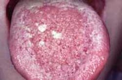
Intraoral examination revealed one diffuse, pebbly lesion located on the dorsal tongue (see photo). The lesion exhibited a vesicular-like surface without ulceration. When palpated, the mass was soft and compressible. Further examination of the oral soft tissues revealed no other lesions present.
Clinical diagnosis
Based on the clinical information presented, which of the following is the most likely diagnosis?
lymphoma
mucocele
hematoma
hemangioma
lymphangioma
Diagnosis
• lymphangioma
Discussion
The lymphangioma is a hamartomatous proliferation of lymphatic vessels. It is a benign tumor that may occur on the skin or mucous membranes. The lymphangioma may involve the oral cavity, head and neck area, legs, arms, or trunk.
Clinical features
Typically, the lymphangioma is a congenital lesion; the term congenital means present at birth. In some instances, it is not present at birth and develops early in life, usually by the age of two. There is no sex predilection.
When found in the oral cavity, the lymphangioma is most often seen on the tongue, palate, buccal mucosa, and lips. The lesion usually appears as a pink or pinkish-red sessile mass. The surface exhibits a characteristic pebbly blister-like appearance and is said to resemble that of tapioca or caviar. The clear blister-like swellings contain lymphatic fluid. When palpated, the lymphangioma is painless and feels soft and compressible.
Diagnosis
The clinical appearance of the lymphangioma is highly suggestive. The lymphangioma may clinically resemble the hemangioma, if there is significant involvement of capillaries; an hemangioma appears reddish-blue or purple.
A biopsy and histologic examination of the lesion is necessary to establish a definitive diagnosis. When examined histologically, the lymphangioma exhibits numerous dilated lymphatic vessels adjacent to the surface epithelium; lymph is found within the vessels.
Treatment
The lymphangioma is a benign lymphatic lesion; this lesion is not contagious and does not have malignant potential. The lymphangioma does not spontaneously regress or disappear with time. The treatment of choice for the lymphangioma is surgical excision. If not completely removed, the lymphangioma may recur.
Joen Iannucci Haring, DDS, MS, is a professor of clinical dentistry, Section of Primary Care, The Ohio State University College of Dentistry.