Case #3
By Joen Iannucci Haring
A seven-year-old female visited a dental office for evaluation of a large swelling on the floor of the mouth.
History
The patient first noticed the swelling two weeks earlier. The patient's mother described the lesion as slowly enlarging over a period of several days, and, since that time, no increase or decrease in size was noted. The patient did not report pain and denied any history of trauma to the area.
The patient appeared to be in a general good state of health with no significant medical history. Her dental history included routine checkups.
Examinations
The patient's vital signs were all found to be within normal limits. Extraoral examination of the head and neck region revealed no enlarged or palpable lymph nodes.
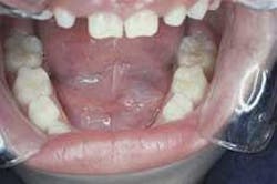
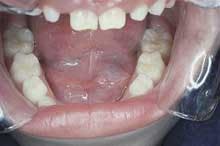
Intraoral examination revealed a well-defined, bluish dome-shaped swelling on the floor of the mouth (see photo). The lesion measured approximately 25mm in diameter and did not cross the midline. Palpation revealed a soft, fluctuant mass. No other soft tissue abnormalities were noted.
Clinical diagnosis
Based on the clinical information presented, which one of the following is the most likely diagnosis?
• dermoid cyst
• pleomorphic adenoma
• mucoepidermoid carcinoma
• hemangioma
• ranula
Diagnosis
• ranula
Discussion
A ranula is a term that is used for a large mucocele that forms on the floor of the mouth. A mucocele can be defined as an accumulation of mucous that results from the rupture of a salivary gland duct and spillage of mucin into the surrounding tissues.
The term ranula is derived from the Latin word rana, meaning frog as this lesion is said to resemble the translucent underbelly of a frog. A ranula is always seen on the floor of the mouth and is often associated with the sublingual gland, but may also be associated with the submandibular or minor salivary glands located in this area.
A ranula is formed when mucous is forced out of a salivary gland duct into the surrounding tissue. Following this pooling of mucous in connective tissue, an inflammatory type response occurs and a wall of granulation tissue is formed.
A ranula is typically seen following trauma of a salivary gland duct, although there is no known history of trauma in many cases.
Clinical features
The ranula may be seen in patients of any age group. The typical presentation is that of a unilateral, dome-shaped swelling on the floor of the mouth. The surface of the lesion is smooth, and the color may vary from pinkish-red to a translucent blue. The lesions that are located close to the surface epithelium are more likely to appear bluish.
The ranula grows slowly and is painless. The size may vary from small to very large; the large lesions may result in displacement of the tongue. Upon palpation, the ranula feels compressible, soft, and fluctuant. A ranula may have a history of rupture and collapse followed by recurrence.
Diagnosis and treatment
The location and clinical features of the ranula are fairly characteristic. The histologic examination of the tissue reveals a lesion similar to a mucocele in other locations. The spilled mucin is surrounded by a granulation tissue response and often contains histocytes.
Treatment of the ranula consists of marsupialization and/or removal of the feeding sublingual gland. Marsupialization is a procedure where the roof of the lesion is removed in order to allow the ducts to re-establish communication with the oral cavity. This procedure is not often successful, in which case a ranula may recur and removal of the offending gland is necessary.
Joen Iannucci Haring, DDS, MS, is a professor of clinical dentistry, Section of Primary Care, The Ohio State University College of Dentistry.

