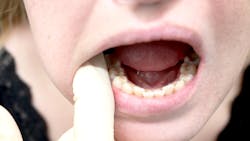
Extra- and intraoral tips to screen for oral cancer
There are an estimated 54,540 new cases of oral cavity or oropharyngeal cancer diagnosed every year.1 No one is better suited to screen for these cancers than dental professionals, who conduct extra- and intraoral screenings every six months. Color changes in tissue, extra- or intraoral lesions, overgrowth of bone, and areas of edema may be indicative of oral cancer. There is also the possibility these changes could be signs of other cancers that have metastasized to the oral cavity.
Here's what dental professionals should look for when doing extra- and intraoral screenings.
Health history review
While the health history review is not conducted intraorally, it’s the first step in conducting a cancer screening as it reveals any conditions that may predispose someone to cancer, oral or otherwise. Tobacco use is a major risk factor and increases a patient's susceptibility by 15-30 times than that of someone who does not use tobacco.2
If a clinician sees any kind of tobacco use on a health history form, they should pay close attention to the mandibular buccal vestibule (dip tobacco), as well as any areas of erythroplakia or leukoplakia as these can indicate oral squamous cell carcinoma (OSCC), the most common oral cancer of the oral cavity.3
Another big risk factor is human papillomavirus. HPV is the most common STD in the United States, accounting for roughly 70% of oropharyngeal cancer cases.4 If a patient has HPV, clinicians should pay special attention to any swelling of the lymph nodes and check the base of the tongue and tonsils for any exophytic lesions. A health history can also help rule out any variants from normal. For example, patients with oral habits such as clenching or grinding may have frictional hyperkeratosis on their buccal mucosa or the lateral margins of their tongue.
Extraoral signs of oral cancer
After thoroughly assessing the health history, the clinician can conduct the extraoral exam. One of the most common areas to detect oral cancer is the vermilion border of the lips. Lip cancer is the second most common malignancy of the head and neck, and 90% of cases are on the lower lip as the upper lip is shielded by the nose.5 While some patients present with dry, chapped lips at every appointment, a key difference between dry lips and potential OSCC is persistent wounds or blisters on the lips that won't heal.5
If a patient consistently has chapped lips, the hygienist should take extraoral photos and recheck the lips for changes in color, texture, or size during subsequent appointments to rule out OSCC. OSCC is "the most common oral malignancy, representing 80% to 90% of all malignant neoplasms of the oral cavity."6 While there has been significant progress in the treatment of OSCC with radiotherapy and chemotherapy, the prognosis is still poor due to metastasis and aggressive local invasion, making early detection and screening paramount.6
Intraoral signs of oral cancer
More than 90% of malignant tumors diagnosed in the oral cavity are OSCC, which is often located on the tongue.7 Most cases present clinically on the lateral border of the tongue, and it’s very rarely seen on the dorsum.8 When checking the lateral border of the tongue, clinicians should check for erythroplakia, leukoplakia, ulcers that won't heal, and any pronounced lesions (raised or flat).8
Many patients also report constant sore throats and may present with bilateral lymphadenitis that can be palpated during the extraoral exam.8 Risk factors for OSCC that should be noted on the patient's health history include smoking, alcohol use, HPV, a familial history of OSCC, and male gender. OSCC has a 2:1 predilection for males.9 A history of any other kind of cancer is also a risk factor as it can metastasize to the oral cavity.9
It can be tough for a clinician to differentiate signs of oral cancer from typical ulcerations such as tongue burns or canker sores. A helpful tip is that canker sores tend to be erythemic and irritated due to localized inflammation, while cancerous lesions typically are not.10 Additionally, canker sores are usually flat and cancerous lesions with lumps or bumps that can be felt upon palpation.10
OSCC is often asymptomatic in its earliest stages, making it difficult for patients to know how long they've had an intraoral lesion. Because canker sores are painful, patients have a much better idea of how long they've had one or what triggered it. Clinicians should always err on the side of caution and refer patients for a biopsy if they aren’t sure about a lesion. Catching OSCC in its earliest stages will have the best prognosis for the patient.
The extra- and intraoral exam is key to diagnosing oral cancer. Most oral cancers in their early stages are asymptomatic and present clinically as other common intraoral lesions, such as canker sores, aphthous ulcers, and tissue trauma. This is why the dental hygienist must thoroughly review the patient's health history, give extra- and intraoral exams every visit, and refer patients for biopsies if any suspicions arise. The only way to catch cancer in its earliest stages is to be hypervigilant when it comes to patient care and oral assessment. Fortunately, these are all strengths of dental hygienists.
References
- HPV and oropharyngeal cancer. Centers for Disease Control and Prevention. October 3, 2022. Accessed March 16, 2023. https://www.cdc.gov/cancer/hpv/basic_info/hpv_oropharyngeal.htm
- Jiang X, Wu J, Wang J, Huang R. Tobacco and oral squamous cell carcinoma: A review of carcinogenic pathways. Tob Induc Dis. 2019;17(4):29. doi:10.18332/tid/105844
- Markopoulos AK. Current aspects on oral squamous cell carcinoma. Open Dent J. 2012;6:126-130. doi:10.2174/1874210601206010126
- McGovern Medical School. Lip Cancer. Otorhinolaryngology–Head & Neck Surgery. December 22, 2022. Accessed March 23, 2023. https://med.uth.edu/orl/2009/12/11/lip-cancer/
- Migueláñez-Medrán BC, Pozo-Kreilinger JJ, Cebrián-Carretero JL, Martínez-García MA, López-Sánchez AF. Oral squamous cell carcinoma of the tongue: Histological risk assessment. A pilot study. Med Oral Patol Oral Cir Bucal. 2019;24(5):e603-e609. doi:4317/medoral.23011
- Okubo M, Iwai T, Nakashima H, et al. Squamous cell carcinoma of the tongue dorsum: Incidence and treatment considerations. Indian J Otolaryngol Head Neck Surg. 2017;69(1):6-10. doi:1007/s12070-016-0979-z
- Oral cavity and oropharyngeal cancer key statistics 2021. Oral Cavity & Oropharyngeal Cancer Key Statistics 2021. Accessed March 16, 2023. https://www.cancer.org/cancer/oral-cavity-and-oropharyngeal-cancer/about/key-statistics
- Pires FR, Ramos AB, Oliveira JB, Tavares AS, Luz PS, Santos TC. Oral squamous cell carcinoma: clinicopathological features from 346 cases from a single oral pathology service during 8 years. J Appl Oral Sci. 2013;21(5):460-467. doi:1590/1679-775720130317
- Types of lip and oral (mouth) cancer. Pennmedicine.org. Accessed March 25, 2023. https://www.pennmedicine.org/cancer/types-of-cancer/mouth-cancer/types-of-mouth-cancer.
- Wang B, Zhang S, Yue K, Wang XD. The recurrence and survival of oral squamous cell carcinoma: a report of 275 cases. Chin J Cancer. 2013;32(11):614-618. doi:10.5732/cjc.012.10219
About the Author

Esmy Ornelas, MS, RDH, CDA
Esmy Ornelas, MS, RDH, CDA, is a professor, clinician, and writer in Oklahoma City. She has 10 years of experience in dentistry and is focused on exploring the professional development aspects of dental hygiene. She can be reached at [email protected].
