Case #7
by Joen Iannucci Haring
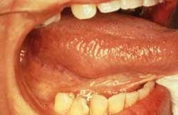
A 46-year-old female visited a dental office for a routine check-up. During the oral examination, a swelling was noted on the lateral tongue.
History
The patient was unaware of the mass on the lateral tongue and denied any pain or sensitivity associated with the swelling. The patient reported no history of trauma to the affected area.
At the time of the dental appointment, the patient appeared to be in an overall good state of health. The patient's medical history was reviewed and it included an allergy to codeine. No significant health problems were noted and no medications were being taken by the patient at the time of the dental examination. The patient's past dental history included sporadic dental examinations, extractions, and routine restorative dental treatment.
Examinations
The patient's vital signs were all found to be within normal limits. Examination of the head and neck region revealed no enlarged or palpable lymph nodes. No significant or unusual findings were discovered during the extraoral examination.
Intraoral examination revealed a large swelling on the right lateral tongue (see photo). Palpation of the area revealed a soft, well-circumscribed nodule that measured approximately two centimeters in diameter. The mass was described as non-painful when compressed. Further oral examination revealed no other lesions present.
The patient was referred to an oral surgeon for an excisional biopsy. Histologic examination revealed an encapsulated tumor consisting of spindle cells organized in whorls and waves surrounding an acellular zone.
Clinical diagnosis
Based on the information available, which of the following is most likely diagnosis?
o traumatic fibroma
o neurilemoma
o neurofibroma
o traumatic neuroma
o malignant schwannoma
Diagnosis
• neurilemoma
Discussion
The neurilemoma (also known as schwannoma) is a relatively uncommon benign tumor of Schwann cells (as the term schwannoma suggests, schwann = Schwann cells; oma = tumor). Schwann cells are found in the neurilemma (outer myelin sheath) of peripheral nerves, and the neurilemoma is derived from the proliferation of these cells. As the proliferation of cells continues, the nerve is pushed aside and is not included in the tumor.
Clinical features
The neurilemoma is most often seen in young- and middle-aged adults. There is no sex predilection. The neurilemoma is typically slow growing and, at discovery, has been present for a long time. The neurilemoma occurs most often as a soft tissue lump of varying size.
When palpated, the neurilemoma feels compressible, well-circumscribed, and movable. The neurilemoma most often occurs in the tongue. The neurilemoma may also occur on the palate, floor of the mouth, buccal mucosa, gingiva, upper lip, and vestibule. The neurilemoma is a benign lesion and does not undergo malignant transformation.
Despite the fact that this tumor arises from nerve tissue, the neurilemoma is typically painless. In instances where the mass compresses an adjacent nerve, rather than the nerve of origin, pain may be a feature. In addition to occurring in soft tissue, the neurilemoma may also occur within bone. In such instances, the neurilemoma most often occurs in the mandible and arises from mandibular nerve tissue. A neurilemoma within bone appears as a unilocular or multiocular radiolucency on a dental radiograph and may cause pain or paresthesia.
Diagnosis
There are no distinctive clinical features of the neurilemoma that enable the practitioner to identify this lesion based on clinical information alone. The diagnosis of a neurilemoma is based on biopsy and histologic examination. The neurilemoma has a characteristic histologic appearance and is seldom confused with other lesions. When examined histologically, the neurilemoma appears as a mass of spindle-shaped cells surrounded by a capsule. There are two microscopic patterns of tissue that may be seen, termed Antoni A and Antoni B.
Treatment
The neurilemoma does not disappear spontaneously or regress with time. The treatment of choice for the neurilemoma is surgical excision. Recurrence of this lesion is unlikely and the prognosis is excellent.
Joen Iannucci Haring, DDS, MS, is a professor of clinical dentistry, Section of Primary Care, The Ohio State University College of Dentistry.

