Case #6

A 39-year-old male presented to a dentist's office for the evaluation of a gingival growth.
History
The patient first noticed the pinkish-red growth near his upper molar five weeks earlier. The patient claimed that the lesion appeared as a small lump and had gradually increased in size. When questioned about any change in color or texture, the patient stated that he was unaware of any such changes. When questioned about any signs or symptoms associated with the growth, the patient stated that the lesion was painless. The patient reported no history of trauma to the affected area.
At the time of the dental appointment, the patient appeared to be in an overall good state of health with no significant past medical history. At the time of the dental visit, the patient was not taking medications of any kind. The patient's past dental history included sporadic dental examinations and routine dental treatment. The patient's last dental appointment was 13 months ago for a dental prophylaxis.
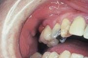
Examinations
Physical examination of the head and neck region revealed no enlarged or palpable lymph nodes. The patient's vital signs were all found to be within normal limits. No significant extraoral findings were noted.
Intraoral examination revealed an elevated reddish-pink nodule located on the gingiva above the cervical region of tooth #3. The lesion appeared as a sessile mass that measured approximately one centimeter in diameter. The lesion exhibited a smooth surface without ulceration or hemorrhage. The mass could be described as well-defined and slightly firm to palpation. The tooth adjacent to the lesion was pulp tested and determined to be vital. Further examination of the oral soft tissues revealed no other masses present.
The patient was referred to an oral surgeon for a biopsy of the lesion. Histologic examination revealed numerous multinucleated giant cells within a background of mesenchymal cells.
Clinical diagnosis
Based on the clinical and histologic information presented, which of the following is the most likely diagnosis?
o peripheral ossifying fibroma
o peripheral giant cell granuloma
o pyogenic granuloma
o parulis
o mucocele
Diagnosis
• peripheral giant cell granuloma
Discussion
The peripheral giant cell granuloma (PGCG) is relatively common and can be described as a reactive lesion that is seen in response to irritation or injury of gingival tissues. In instances where trauma is believed to be a precipitating factor, a history of tooth extraction, denture irritation, or a chronic infection may be significant. The PGCG is believed to arise from the periodontal ligament or the periosteum.
Clinical features
Although the PGCG may be seen at any age, most cases occur between the ages of 40 and 60. Females are affected more frequently than males. The PGCG is found exclusively on the gingival and alveolar process. It usually occurs between the first molars and incisors; the mandibular anterior gingiva is most often affected. The lesion usually appears as a red or blue broad-based or "sessile" mass. The surface may be smooth or granular and may exhibit ulceration if traumatized. The PGCG will feel slightly firm to palpation and usually measures less than two centimeters in diameter.
In some cases, the PGCG may cause bone resorption of the underlying bone. When the PGCG is seen in an edentulous area, a superficial erosion or "cupping out" of bone may be identified on a corresponding radiograph.
Diagnosis
The PGCG may clinically resemble the pyogenic granuloma, the mucocele, or the peripheral ossifying fibroma. A biopsy and histologic examination of the tissue is necessary in order to make a definitive diagnosis. When examined histologically, the PGCG exhibits a hyperplasia of fibroblasts with scattered abundant multinucleated giant cells. The multinucleated giant cells may exhibit up to several dozen nuclei.
Treatment
The PGCG is a reactive lesion. This lesion is not infectious and does not have malignant potential. The PGCG does not spontaneously disappear or regress with time. The treatment of choice is surgical excision. The surgery should include the base of the lesion and extend to the periosteum of bone. The curettage of the underlying bone is recommended along with removal of local factors, such as calculus on the adjacent teeth. About 10 percent of these lesions recur after removal.
Joen Iannucci Haring, DDS, MS, is a professor of clinical dentistry, Section of Primary Care, The Ohio State University College of Dentistry.