Eye spy: What the eyes can tell us about the mouth
Author’s note: All of the patients depicted in this article’s photos had low tongue postures, retrognathic maxillae, and chronic airway issues.
Whoever said that “The eyes are the window to the soul” clearly wasn’t an orofacial myologist. If that person had any inkling to what the eyes can tell those of us who specialize in correcting oral rest posture for a living, the saying would be, “The eyes are the window to the health of your upper airway,” or perhaps “The eyes are the window to your underdeveloped maxilla.” And so, while the rest of the world is gazing into one another’s eyes, searching for the secrets of the soul, I’ll be over here teaching my fellow dental professionals how to look for the subtle clues that the eyes can give us about the health of our patients before we ever take a look inside of their mouths.
As a whole, dentistry has made enormous advancements in connecting the dots between airway issues, sleep, and the oral cavity. We are doing a much better job of recognizing the red flags, such as large tonsils and chronic mouth breathing, and then questioning our patients about the quality of their sleep. But what about the not-so-obvious cases? What about the patients who present with much more subtle signs of trouble? Not every patient will present with a gaping mouth and kissing tonsils. If we learn to look at the eyes of our patients, they may tell a story that is not readily apparent on their medical history.
Clue no. 1: Dark circles
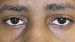
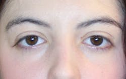
The first and most common clue is periorbital hyperpigmentation, commonly known as “allergic shiners” (figures 1 and 2). Allergic shiners are the dark bruise-like circles that many of our pediatric and adult patients have under their eyes. We all experience darkening under the eyes from time to time, especially during a cold or the flu, or after too many late nights in a row. Acute darkening under the eyes is typically not a concern when it resolves once we feel better or catch up on sleep. But what about our patients who have chronic darkness, puffiness, or redness around their eyes? Dark circles can tell us a lot about a patient’s airway health.
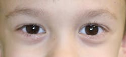
As you have probably already deduced from the name, the most common cause of allergic shiners is … you guessed it—allergies! Stuffy, congested nasal passages lead to congestion in the small blood vessels under the eyes. This swelling of the soft tissue causes venous pooling, or poor drainage of the blood vessels, which causes darkness and puffiness to appear.1 If a patient suffers from seasonal allergies, allergic shiners can come and go with the seasons. However, this darkening will remain year-round if the allergy is related to food; indoor allergens such as pets, mold, or dust mites; or irritants such as cigarette smoke or other pollutants in the air. Allergic shiners can also be a sign of enlarged adenoids, nasal polyps, or sleep apnea.1–4
When our patients have chronic airway issues, we need to dig a little deeper and observe how their stuffy noses are affecting their oral rest posture. For proper growth of orofacial complex to occur, the tongue needs to rest on the palate when it isn’t in use. In addition to proper tongue posture, the lips should be gently sealed and the jaws should be no more than 2 mm to 3 mm apart.2,3 Patients with chronic airway issues, such as allergies, will develop habitual mouth breathing. Mouth breathing changes the oral rest posture and leads to narrowing of the jaws, which in turn leads to narrowing of the nasal cavity.2–5 In dentistry, we tend to view the maxilla as an oral structure, but in reality, only half of the maxilla is in the oral cavity. The other half makes up the floor of the sinus cavity. Those allergic shiners may be the first clue to a chronic airway problem that causes narrow jaws and crowded teeth.
Narrowing of the maxilla due to poor tongue posture from mouth breathing also leads to narrowing of the spaces behind the maxilla, specifically the pterygopalatine and infratemporal fossae.5–7 These bony spaces contain the pterygoid venous plexus, which receives blood from the inferior orbital vein. When the fossa space is reduced, the pterygoid venous plexus shrinks, pushing the venous blood back through the inferior orbital vein.5 In this situation, the venous pooling is caused not just by inflammation of soft tissues, but by change in the structure from years of improper breathing and oral posture.
Dark circles below the eyes can lead us to investigate the patency of the nasal airway and to look for poor oral rest posture and narrowing of the maxilla. Appropriate referrals for this type of patient would be an allergist or ENT specialist to address the nasal airway and an orthodontist to address palatal expansion. An orofacial myologist can help reestablish a proper oral rest posture and help establish nasal breathing.2–4
Clue no. 2: Visible sclera under the iris
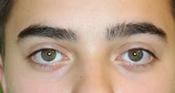
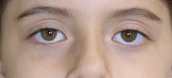
The second clue that we can look for in the eyes of our patients is visible sclera (the white part of the eye) below the colored iris (figures 3 and 4). No sclera should be showing above or below the iris,8 but we tend to see “sleepy eyes” in some patients. If we consider that the maxilla makes up a large portion of the orbit of the eye, it’s easy to see how we can spot midfacial hypoplasia before looking inside of a patient’s mouth. Patients with hypoplasia of the midface also have flatter faces and typically suffer from the numerous symptoms of sleep-disordered breathing, which can include allergic shiners and puffy eyes, as well as a narrow dolichocephalic face.2,3,5,8 These patients will benefit from an ENT referral for an airway assessment as well as a referral for orthodontic treatment. Depending on the severity of the midfacial hypoplasia, they may require surgical advancement of the jaw(s) in conjunction with orthodontic treatment.9 An orofacial myologist can help improve oral rest posture and function during treatment of the midface deficiency.
Clue no. 3: Downturned canthi
Downturned lateral canthi are another subtle clue that the maxilla has not grown correctly (figure 5). When the lateral canthi of the eyes are lower than the medial canthi, the patient’s eyes will have a “droopy” appearance.5,9 A wide, well-positioned maxilla gives support to the orbit of the eyes, but it also supports the soft tissue around the eyes.10 The proper position of the maxilla can help lift the eyes and give the cheekbones a full, healthy look. A droopy-eyed appearance indicates maxillary insufficiency.5 As mentioned before, we need to dig deeper and understand how craniofacial bone growth patterns can give clinicians important clues about patients’ oral rest postures. Anytime that we see signs of maxillary hypoplasia, we need to investigate the rest posture of the tongue, jaws, and lips, and closely evaluate the patient’s breathing patterns. When airway interferences are present or sleep is affected, we need to make the appropriate referrals to an allergist or ENT specialist and possibly a separate physician who specializes in sleep. An orthodontist can address the structural deficits, and an orofacial myologist can work to improve the oral rest posture and address abnormal function and breathing.
Conclusion
When we stop and consider that bone structure creates the framework and attachments for soft tissue, it is easier to see how structure, function, and appearance go hand in hand. We can tell a lot by looking at the faces of our patients. The eyes really are the window to so much more than the soul. They are the window to the health of the airway and the window to the bone structure of the oral cavity and facial skeleton. It’s time to start looking at more than just the oral cavity—it’s time to treat the whole person attached to those teeth!
References
1. Allergic shiners: Symptoms, causes, and treatment. Healthline website. https://www.healthline.com/health/allergic-shiners. Published May 4, 2017. Updated August 28, 2018. Accessed July 15, 2019.
2. Gelb M, Hindin H. Gasp! Airway Health: The Hidden Path to Wellness. CreateSpace Independent Publishing Platform; 2016.
3. Kahn S, Ehrlich PR. Jaws: The Story of a Hidden Epidemic. Stanford, CA: Stanford University Press; 2018.
4. Schoem SR, Darrow DH. Pediatric Otolaryngology. Washington, DC: American Academy of Pediatrics; 2012.
5. Liem E, ed. Sleep Disorders in Pediatric Dentistry: Clinical Guide On Diagnosis and Management. Cham, Switzerland: Springer Nature; 2019.
6. Lopez D. How swallowing incorrectly can affect your eyes. Daniel Lopez, DO website. https://www.daniellopezdo.com/swallowing-incorrectly-can-affect-eyes. Published July 3, 2017.
7. Najeeb DN. Pterygopalatine Fossa - Gross Anatomy. YouTube. https://www.youtube.com/watch?v=2fxF0z5DdB8. Published October 13, 2014.
8. Moore S. Sleep Wrecked Kids: Helping parents raise happy, healthy kids, one sleep at a time. New York, NY: Morgan James Publishing; 2020.
9. Soydan SS, Bayram B, Sar C, Uckan S. Change in inferior sclera exposure following Le Fort I osteotomy in patients with midfacial retrognathia. J Oral Maxillofac Surg. 2014;72(1):166. doi:10.1016/j.joms.2013.09.025.
10. Boshart CA. The Myofunctional Evaluation. 3rd ed. Ellijay, GA: Speech Dynamics; 2017.
Angie Lehman, RDH, COM, has been certified by the International Association of Orofacial Myology (IAOM) and has practiced orofacial myology exclusively since 2012. She currently serves on the board of directors for the IAOM. Lehman is owner of Oral Myofunctional Therapy of York, a private practice in York, Pennsylvania, that provides myofunctional therapy to children and adults. She also provides continuing education for dental and medical professionals. Her passion is to see dental and medical professionals work collaboratively to better understand oral function and craniofacial development and to incorporate therapy into their specific areas of care.
About the Author

Angie Lehman, RDH, COM
Angie Lehman, RDH, COM, has been certified by the International Association of Orofacial Myology (IAOM) and has practiced orofacial myology exclusively since 2012. She currently serves on the board of directors for the IAOM. Lehman is owner of Oral Myofunctional Therapy of York, a private practice in York, Pennsylvania, that provides myofunctional therapy to children and adults. She also provides continuing education for dental and medical professionals. Her passion is to see dental and medical professionals work collaboratively to better understand oral function and craniofacial development and to incorporate therapy into their specific areas of care.